2.1 Structures and Properties of Amino Acids
2.1.1 Structure of amino acids
An amino acid is a monomer that contains a central carbon atom (see figure 2.3). It can be joined via peptide bonds; there are 20 known amino acids.
Some amino acids may appear more frequently than others (e.g., methionine) while some don’t appear very often (e.g., proline and its derivatives)!

Figure 2.3: Structure of an Amino Acid
Every amino acid is also bound to…
- an amino group.
- a carboxyl group.
- a hydrogen atom.
- a side chain (i.e., the “R” group).
Different amino acids are distinguished by their “R” groups!
2.1.2 20 common amino acids and their pKa values
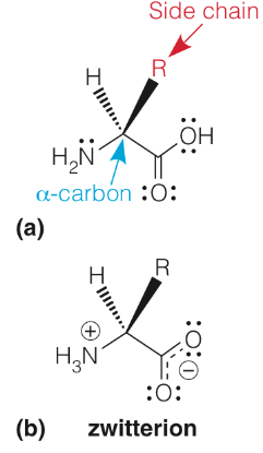
Figure 2.4: Acidic and Basic Components of Amino Acids
The pKa of an amino acid’s carboxylic group is about 2; the pKa of its amino group is about 10. Hence, when the amino acid in figure 2.4 is present at physiological pH (i.e., pH 7), both the amino and the carboxylic group will be ionized to yield the zwitterion form.
Because of this, amino acids are also typically written in their zwitterionic forms!
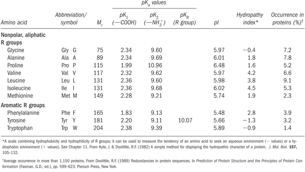
Figure 2.5: pKa Values of 20 Common Amino Acids
Figure 2.5 shows the pKa values of 20 common amino acids. The amino acids histidine and cysteine are unique in that their side chain pKas are closer to physiological pH. Because of this, these amino acids also play a special role in enzymology as the functional group of catalysts.
2.1.2.1 Non-polar, aliphatic amino acids
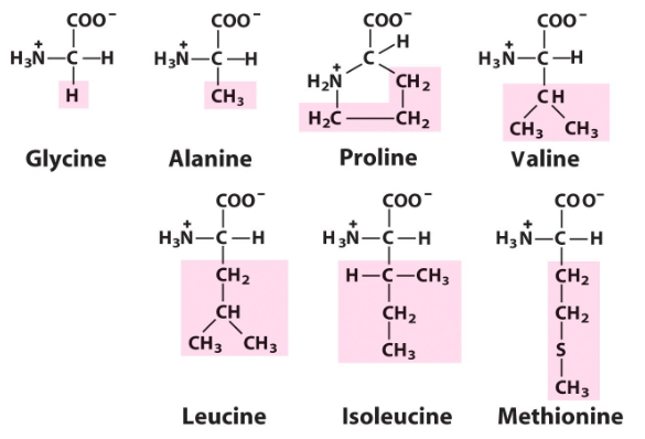
Figure 2.6: Nonpolar, Aliphatic Amino Acids
These amino acids are usually found within the core of a protein (where they are shielded from water).
2.1.2.2 Polar, uncharged amino acids
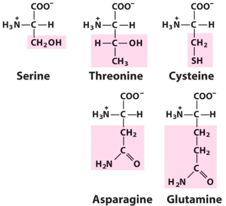
Figure 2.7: Polar, Uncharged Amino Acids
Figure 2.7 displays five polar and uncharged amino acids, all of which are capable of forming hydrogen bonds with other compounds. The OH group of serine and the SH group of the cysteine are great nucleophiles and play key roles in enzymatic activity.
Unlike their acidic counterparts, the amino acids asparagine and glutamine have uncharged polar side chains too!
2.1.2.3 Positively charged amino acids
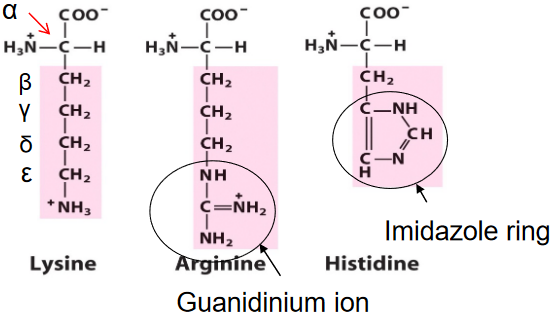
Figure 2.8: Positively Charged Amino Acids
The amino acids lysine (pKa = 10.5) and arginine (pKa = 12.5) are the most basic amino acids. Their side chains are almost always positively charged at physiological pH.
The guanidinium ion in arginine (circled in figure 2.8 ) makes the amino acid the most basic because of the resonance stabilization of the protonated side chain.
2.1.2.4 Negatively charged amino acids
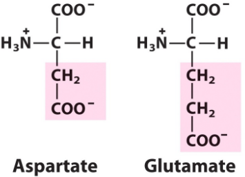
Figure 2.9: Negatively Charged Amino Acids
Aspartic acid (pKR = 3.7) and glutamic acid (pKR = 4.3) generally carry negative charges at physiological pH. These amino acids are also referred to as aspartate and glutamate (i.e., their conjugate bases instead of their acids - this is shown in figure 2.9 ).
These amino acids also play important roles in electrostatic interactions and metal ion (e.g., Mg2+ and Ca2+) binding.
2.1.2.5 Aromatic amino acids
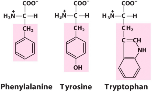
Figure 2.10: Aromatic Amino Acids
As seen in figure 2.10, there are three aromatic amino acids.
Phenylalanine is one of the most hydrophobic amino acids. The amino acids tyrosine and tryptophan also have some hydrophobic character, but it is tempered by the polar groups in their side chains.
2.1.3 Modifications of amino acids
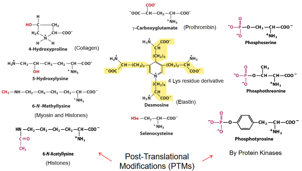
Figure 2.11: Post-Translational Modifications of Proteins
The post-translational modification (some examples are shown in figure 2.11) of proteins has a multitude of functions in the cell: from signal transduction to gene regulation to the modification of dynamics and structure.
2.1.3.1 Disulfide bond formation
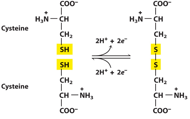
Figure 2.12: Disulfide Bond Formation Between Two Cysteine Residues
A disulfide bond can be formed between two cysteine residues via the oxidation of two cysteine molecules. These disulfide bonds also serve to stabilize the structures of many proteins.
Do also note that disulfide bond formation alters the primary structure of proteins (as the covalent bonding activity is also modified).
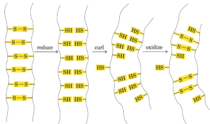
Figure 2.13: Permanent Curls in Hair
The reaction shown in figure 2.12 has also been utilized in the cosmetic industry (though it has fallen out of fashion). In figure 2.13, disulfide bonds are broken and reformed to yield a permanent curl.