2.4 Spectroscopic Properties of Amino Acids
All aromatic amino acids (i.e., phenylalanine, tyrosine, and tryptophan) absorb UV light due to their delocalized electrons:
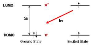
Figure 2.18: Delocalized Electrons Accepting Light
Note that the terms HOMO and LUMO in figure 2.18 stand for highest occupied molecular orbital and lowest unoccupied molecular orbital respectively.
Nonetheless, the absorption3 in figure 2.18 can be used to measure protein concentration via the Beer-Lambert law:
\[\begin{equation} A = \epsilon \times C \times L \tag{2.1} \end{equation}\]
Where:
- \(A\) is the absorbance
- \(\epsilon\) is the molar absorption coefficient
- \(C\) is the concentration of the substance (in this case, the protein)
- \(L\) is the wavelength of the light.
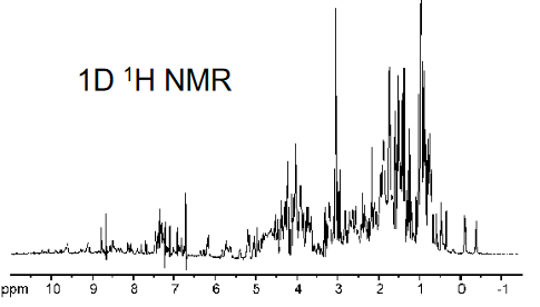
Figure 2.19: 1D H NMR Example
A 1H NMR spectra (shown in figure 2.19) is characteristic of each residue in a protein, and higher resolution NMR measurements can be used to depict the three-dimensional structures of peptides and small proteins.
An absorbance at 280 nm is a good diagnostic device for proteins↩︎