4.1 Determining the 3D Structure of Proteins
There are three ways this can be done:
- X-ray crystallography
- Nuclear magnetic resonance spectroscopy
- Electron microscopy
4.1.1 X-ray crystallography
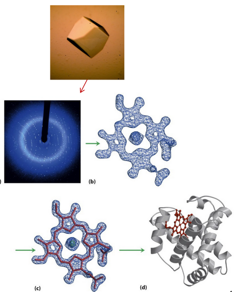
Figure 4.2: Proteins in Various Stages of X-Ray Crystallography
Once the purified protein in question has been crystallized, x-ray crystallography can then be performed on the crystal.
The diffraction pattern of the protein crystal can then be translated to yield a three-dimensional electron density distribution via a Fourier transform.
This map can then be used to build the atomic structure of the protein or nucleic acid molecule (as seen in figure 4.2).
4.1.2 Nuclear magnetic resonance spectroscopy
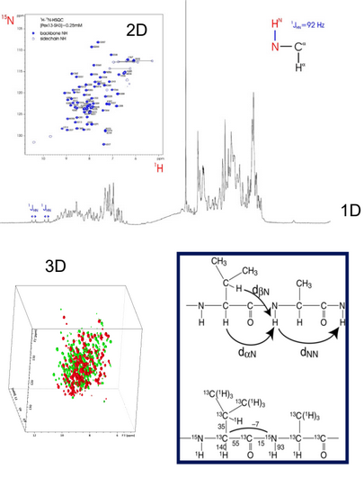
Figure 4.3: Multidimensional NMR
The three-dimensional structure of macromolecules can also be determined in solution by employing multidimensional (i.e., 1D / 2D / 3D - see figure 4.3) NMR analyses of the macromolecule sample.
NMR characterizations can also reveal structures and dynamics.
4.1.3 CATH system
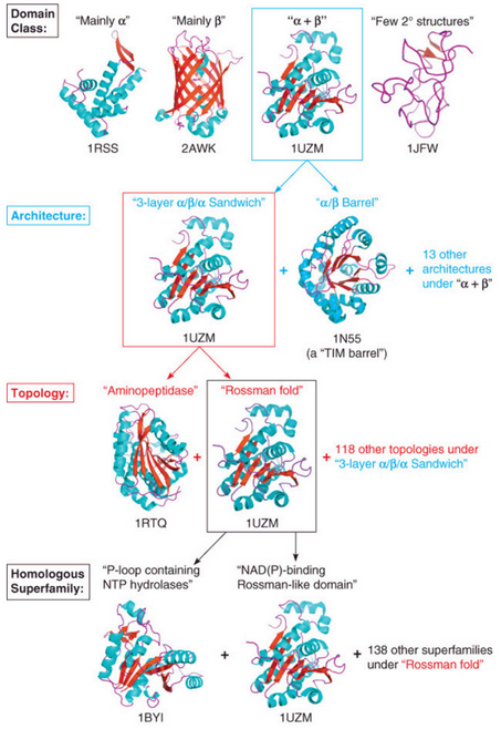
Figure 4.4: CATH System at Play
The CATH system is a classification system (see figure 4.4) based on four protein features:
- Class
- Architecture
- Topology
- Homologous superfamily
There are well over 100000 known protein structures, but groups of these structures share similar features in terms of their overall fold or specific structural attributes.
Note that a similarity in protein fold or structure does not mean that the protein shares an evolutionary relationship with another protein.