6.3 Secondary and Tertiary Structure of DNA
The secondary and tertiary structure of DNA was largely based on Chargaff’s rule and [data from X-ray diffraction] based on studies done by scientists Franklin and Wilkin. In 1953, two scientists Watson and Crick then proposed a model for DNA - their model was called the DNA double helix.
The strands of DNA are anti-parallel to one another and wound around each other as well. The bases of DNA are located on the inner sides of the helix; the sugars and phosphate heads of DNA are located on the outside of DNA to form the backbone of the helical structure of DNA.
6.3.1 Chargaff’s rule
Chargaff’s rule states that the DNA content of “A” is equivalent to the DNA content of “T” and that the DNA content of “G” is equivalent to the DNA content of “C.”
This rule is also explained by Watson-Crick base pairings.
6.3.2 Base pairing
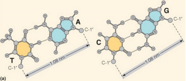
Figure 6.14: Bonding in DNA
Hydrogen bonding occurs between parallel pairs of bases; one base always pairs with another on the anti-parallel strand (see figure 6.14)!
The A-T base pair is stabilized by two hydrogen bonds; the G-C base pair is stabilized by three hydrogen bonds.
6.3.3 Base pair stacking
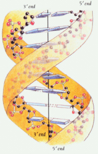
Figure 6.15: Base Pair Interactions
The planes of DNA’s bases are perpendicular to its axis, hence allowing for the stacking of aromatic rings. Base stacking (see figure 6.15) permits van der Waals interactions to take place - these interactions are also the major driving force that stabilizes the double helix (see figure 6.15).
6.3.4 Base pair rotation
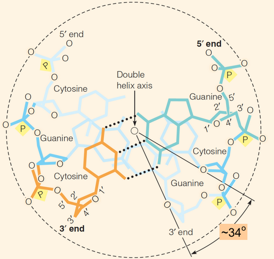
Figure 6.16: Base Pair Rotations
Each base pair in a molecule of DNA is rotated by about 34\(^\omicron\) with respect to the next. This is to accomodate for the next ~10.5 base pairs (i.e., helical repeat) for each turn in the B-form of DNA (see figure 6.16).
6.3.5 Base pair distance
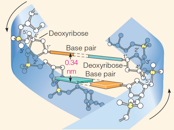
Figure 6.17: Distances Between Base Pairs in DNA
The distance between each base pair (i.e., the rise) is about 3.4 Angstroms. Given the helical repeat of ~10.5 bases per turn, the repeat distance (i.e., the pitch) is then ~36 Angstroms in the B-form of DNA (see figure 6.17).
This base pair separation corresponds closely to the van der Waals thickness of the bases - this is to ensure that the base pairs are closely stacked within the double helix.
6.3.6 Major and minor grooves of DNA
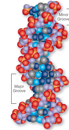
Figure 6.18: Major and Minor Grooves in DNA
There are two exterior grooves between the protruding sugar-phosphate backbones. The major grooves are of different size from the minor grooves (see figure 6.18) because of how the bases in DNA are asymmetrically attached to the pentose sugar.