7.2 Types of Diagnoses
7.2.1 Specimen collection
As a general rule of thumb, one should sample the site of infection.
Swabs used for molecular assays should be compatible with the molecular assay for which they are intended. This is because incorrect sampling equipment can result in false-negative results - wooden shafts are toxic to some bacteria and so are cotton-based swabs (e.g., chlamydiae).
Multiple samples are also encouraged - in the case of blood samples, blood from various areas of the body are often taken to increase the probability of finding the pathogen.
Lastly, pathogens must be transported quickly in the correct medium so as to prevent the growth of other flora. At the very least, the pathogen has to be refrigerated.
7.2.2 Overview of microscopy methods
Most specimens are stained with some sort of dye - different pathogens require differetn stains and no dye is 100% pathogen-specific.
Some commonly used stains are the:
- Gram stain
- Acid fast stain
- Fluorescent stain
- Indian ink stain
While stains can be quick, the accuracy of the stain also depends on the experience of the operator. For this reason, specifities are low, but sensitivities are high for stains.
To counter the above, one can consider coupling an antibody to a dye to increase both specifity and sensitivity.
7.2.3 Cell cultures
A culture is microbial growth on nutritional solid or liquid medium - an increased number of organisms also simplify identification. Cultures also aid in the testing of antimicrobial susceptibility.
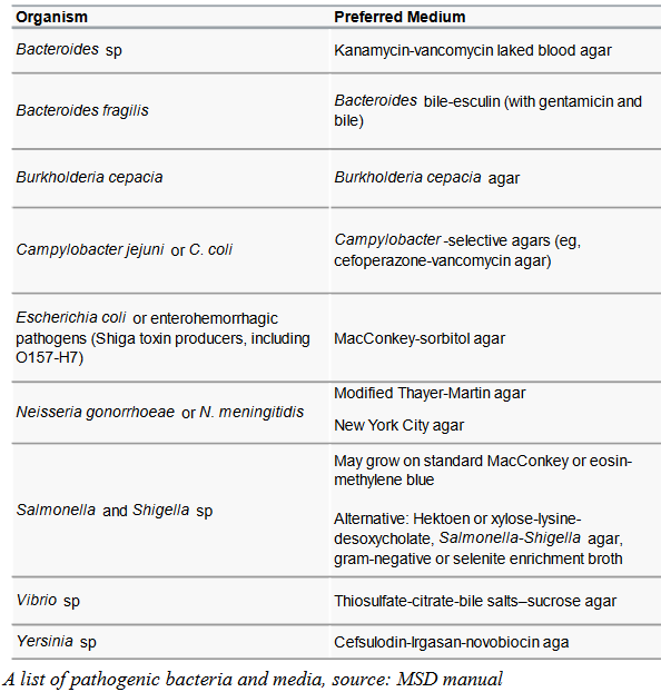
Figure 7.2: Types of Media Used for Varying Pathogens
However, different pathogens all require some sort of specific media and even then, not all pathogens can be cultured. Cultures also take time and are vulnerable to cross contamination.
For this reason, cultures are generally more for confirming diagnoses; while it is possible to cultivate viruses and fungi, these are likely even more difficult than bacteria.
7.2.3.1 Identifying pathogens from a cell culture
Assuming one does not use genetic methods, some possible ways are:
Phenotypes
That is, via the pathogen’s color, shape, and colony size.
Staining
Analysis for metabolite biomarkers
For instance, GC content analysis for fatty acid profiles.
Protein marker analysis with mass spectroscopy
A MALDI-TOF could be used to identify yeasts, bacteria, molds, and viruses.
7.2.4 Immunologic tests
These are generally used to detect a pathogen or an antigen of a pathogen. Serial dilution can be used to measure titre (i.e., quantitation).
These tests are vital to medical science because:
These tests have shortened hospital stays and decreased illness severities by identifying and assessing disease progression.
Immunoassays can be used to identify different proteins, hormones, and antibodies.
Immunoassays can also be used to find contaminants in food and water (hence aiding in the quality control of these products).
7.2.4.1 Some immunologic tests for infectious diseases
Precipitation tests
These measure antibody-antigen complexes via visual perceptions within a gel or a solution.
These tests have low sensitivity and as such, are not very used today.
Agglutination tests
Small particles couple to the antigen or the antibody and is mixed with the pathogen.
This test is fast, but is not very sensitive.
Western blot test
Antibodies in the patient are tested by binding their antigens to a membrane via blotting.
This test provides good sensitivity, is highly specific, and is often used for confirmation purposes.
Complement fixation
An antibody-antigen complex depletes complement proteins that would lyse sheep red blood cells that have been bound to antibodies, hence turning the solution pink.
This test isn’t that used nowadays.
Competitive inhibition immunoassay
We used a labelled analyte to compete with an unlabelled analyte for binding to an antibody. The quality of the labelled analyte is inversely proportional to the concentration of the unlabelled target analyte in the sample.
This test is often used for small analytes since it only requires single antibody-binding.
Enzyme immunoassays
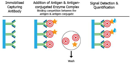
Figure 7.3: How ELISA works
We use antibodies linked to enzymes to find antigens and detect and quantify antibodies - for instance, EIA and ELISA.
This test has high sensitivity and high throughput and is usually used for screening purposes.
Other tests…
Radio-Immunoassay measures antigen-anitbody binding via radioisotopes.
7.2.4.2 ELISA assay formats
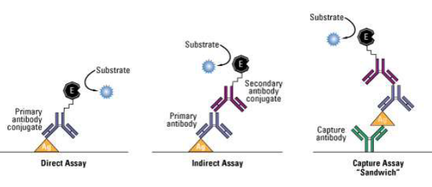
Figure 7.4: A Protein ‘Sandwich’
The antigen of interest can be fixed onto an assay via an antibody that has been attached to the assay.
The antigen is then detected via a labelled primary antibody (i.e., directly) or indirectly (i.e., indirectly) - this then forms a “sandwich”: the most powerful ELISA assay format as it is sensitive and robust.
7.2.5 Antimicrobial susceptibility testing
Susceptibility tests are used to figure out a microbe’s susceptibility to a drug by exposing a standardized concentration of an organism to varying concentrations of antimicrobial drugs. This test is applicable to viruses, bacteria, and fungi.
However, this is an in vitro test and hence, not representitive of in vivo conditions. Yet, this test can also be qualitative, semi-quantitative, and nucleic-acid based.
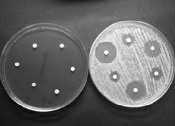
Figure 7.5: Dark Diffusion Method
The dark diffusion method in susceptibility testing is suitable for rapid-growing organisms - the diameter around each zone of inhibition is measured. Here, antibiotic-impregnated disks on agar plates are inoculated with the pathogen.
7.2.5.1 More on susceptibility testing
Semi-quantitative methods
These determine the minimal concentration of a drug that inhibits the growth of the pathogen in vivo.
We use agar / broth or a strip of polyester that have been impregnated with varying concentrations of antibiotics.
Nucleic acid based methods
Here, we find known resistance genes and mutations - however, these methods have issues concerning false positive and false negative results.
These methods are also more useful for detecting multi-drug resistant infections in at-risk groups and can also help find possible resistance in organisms in positive blood cultures.
7.2.5.1.1 Detecting infections based on nucleic acids
We can find organism-specific DNA or RNA sequences in samples with or without amplification via staining, spectrophotometry, radioisotopes, enzymatic reactions, fluorescence, or whole genome sequencing.
Generally speaking, these methods are specific, highly sensitive, and provides rapid results. These methods can also be qualitative or quantitative.
Of course, one can also amplify the amount of genetic material present via:
Target amplification
PCR and RT-PCR
Signal amplification
Branch DNA assays and hybrid capture
Probe amplification
Ligase chain reaction, cleavase invader, and cycling probes
Post-amplification analysis
Sequencing and microarrays
7.2.5.1.2 TaqMan Real time quantitative PCR
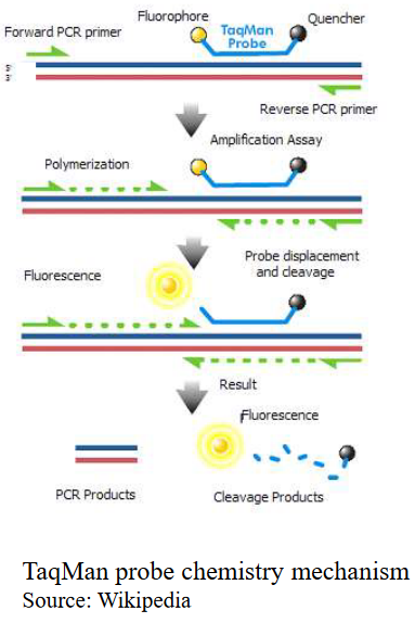
Figure 7.6: A Short Schematic on TaqMan Probe Chemistry
This test was made to improve the specificity of quantitative PCR. The TaqMan probe relies on the principle of 5’ to 3’ exonuclease activity of Taq-polymerase.
The TaqMan probe consists of a fluorophore that has been covalently attached to the 5’ end of the oligonucleotide probe and a quencher at the 3’ end.
When Taq polymerase moves, it then cleaves and frees the fluorophore, hence emitting fluorescence.
This test is used in:
- Finding viral loads of specimens
- Gene expression assays
- DNA quantification
- SNP genotyping