Chapter 11 Microscopy Concepts To Know
11.1 A Brief Introduction to Optics
11.1.1 So what is light?
"Light," as we colloquially refer to it, is a form of electromagnetic radiation that is visible to the human eye. Visible light comprises a small section of the electromagnetic spectrum, encompassing wavelengths from approximately 380-700nm (depending on how good your eyes are!). Because light travels as a wave and interacts as a particle (carrying bundles of energy called photons), we can manipulate many of its properties in order to capture images far beyond what the naked eye can see.
11.1.2 The Wave Nature of Light
For brightfield and confocal microscopy, there are 6 main properties of
light that we will focus on that allow us to create optimized images.
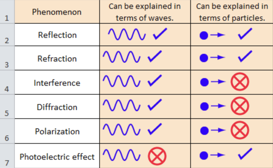
Each phenomena will be described briefly below (with an example).
Reflection: when a ray of light hits a smooth surface and bounces
back (i.e. when you look in a mirror).
Refraction: when a light
wave propagates through a different medium it bends due to the change in
refractive index (i.e. fish that look slightly distorted in water or
the Pink Floyd Dark Side of the Moon album cover).
Interference: occurs when multiple waves interact with each other. Interference can
either be constructive (increases the amplitude of the resultant wave)
or destructive (decreases the amplitude of the resultant wave) (i.e.
when two musicians play a note of the same frequency and it sounds
louder).
Diffraction: occurs when a wave bends to fit through an
aperture (i.e. you can hear someone talking in an adjacent room through
the open doorway because the sound waves diffract).
Polarization: transverse waves can be filtered/blocked based upon their geometry
(i.e. "polarized" sunglasses that block glare).
11.1.3 What does this have to do with microscopes?
Due to the wave properties of light described in the previous section,
specifically interference and diffraction, light from a microscope forms
an Airy disk.
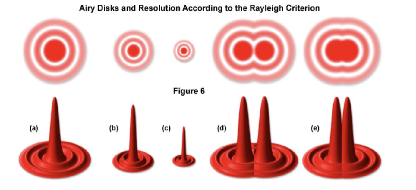
An important criterion for resolution is the Rayleigh Criterion,
which tells us that two objects are considered resolved when the Airy
disk (central maximum) of the first object is directly overlapped with
the first order minimum of the diffraction pattern of the second object.
Our microscope features airyscan, which uses an array of pinhole
apertures and deconvolution to beat the Rayleigh Criterion.
It is important that you choose the correct objective for your sample and desired resolution. The resolving power (R) can be found for a traditional confocal microscope with a given objective using the following formula: \[R_{xy} \approx \frac{0.61 \lambda}{NA},\] where \(\lambda\) is the wavelength of the laser (excitation light) and NA is the numerical aperture of the objective. For a microscope equipped with airyscan, the resolution is twice as good. Due to differences in the Point Spread Function, resolution is poorer in the \(z\)-direction (which is why blocking should be done in the \(xy\)-plane if possible). The resolution in the \(z\)-direction is given by \[R_{z} \approx \frac{2 \lambda n}{(NA)^{2}},\] where \(n\) is the refractive index of the medium in which your sample is fixed.