14.1 Setup of a fMRI
There are several parts to a fMRI machine:
Magnet
This is a large magnetic field that aligns protons in the body over a large area.
Radiofrequency coils
These components transmit and receive radiofrequency into and from the body.
Gradients
This induces a linear change in the magnetic field and performs spatial encoding.
Computer system and consoles
Patient handling system
14.1.1 BOLD in fMRI
fMRI utilizes the BOLD-dependent contrasts. BOLD is short for “blood-oxygen level dependent”.
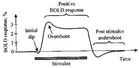
Figure 14.3: BOLD Response in a fMRI
Brain and body scans can be used to map neural activity in the brain or in the spinal cord of humans or other animals by imaging the change in blood flow based on neural activity.
14.1.1.1 BOLD Contrasts
Deoxyhemoglobin (i.e., dHB) is paramagnetic while oxyhemoglobin is not as paramagnetic. This magnetism increases with increasing oxygenation levels.
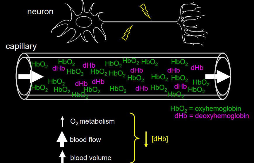
Figure 14.4: Oxyhemoglobin and deoxyhemoglobin in a vein
Venous and capillary blood has a higher proportion of deoxyhemoglobin. Hence, the difference in the oxy and the deoxy states is greater for veins (BOLD is sensitive to such changes).
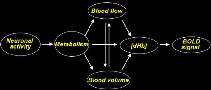
Figure 14.5: Oxyhemoglobin and deoxyhemoglobin in a vein
dHB has a different resonance frequency than water and hence, is used as a contrast agent. Inside blood vessels, dHB creates frequency offsets in surrounding tissues - this spreading of frequency creates a loss in signal over time.
In a sense, dHB is a super indirect measure of activity
14.1.2 Hemodynamic Responses
The hemodynamic response (i.e., HDR) is the change in MR signal from neural activity. It lags neuronal events triggering it by one to two seconds - this is how long it takes for the vascular system to respond to the brain’s need for glucose.
At this point, it takes about 5 seconds for the HDR to go to a peak. If neurons continue to fire, then the peak plateaus while the neurons stay active.
The BOLD signal falls below its original levels after activity has stopped; the signal then recovers to a baseline. There is also some evidence that continuous metabolic requirements in brain regions contribute to this.