5.2 Confocal Microscopes
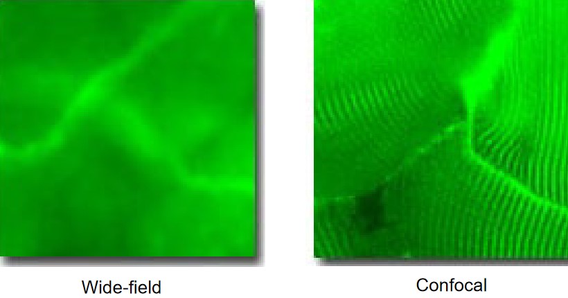
Figure 5.4: Rabbit Muscle Cells Viewed Under Wide-Field Fluorescence Microscopy
During image viewing, only the focal plane is in focus, but all fluorophores that are illuminated contribute to the final image.
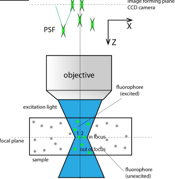
Figure 5.5: Focal Planes in Fluorescence Microscopy
Fluorophores (i.e., ‘1’ and ‘2’ in the above figure) within the focal plane are the objects of interest and are focused as the final image as PSF 1’ and PSF 2’ respectively.
However, fluorophores outside the focal plane (i.e., the out-of-focus blur) may also contribute to the final image via their PSF projections. The larger an image plane away from a PSF’s center, the larger and the weaker the PSF projection.
The resulting image has background haze when all the PSFs have been summed up.
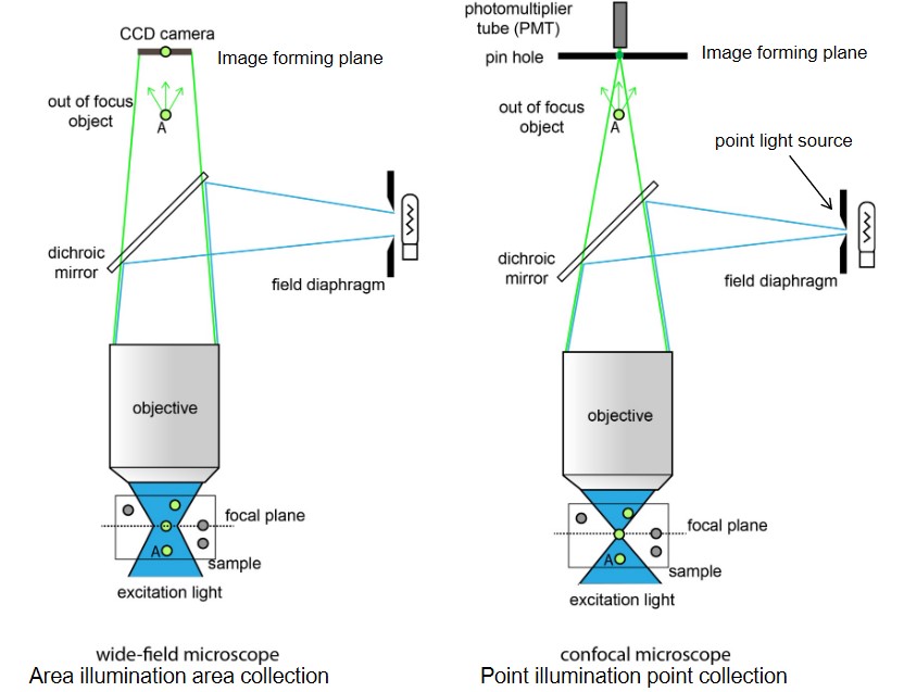
Figure 5.6: Confocal Microscopes vs. Wide-Field Microscopes
Confocal microscopes can reduce out-of-focus blurs. This is because confocal microscopes use lasers and pinholes to prevent out-of-focus lights from reachign the photomultipler tube.
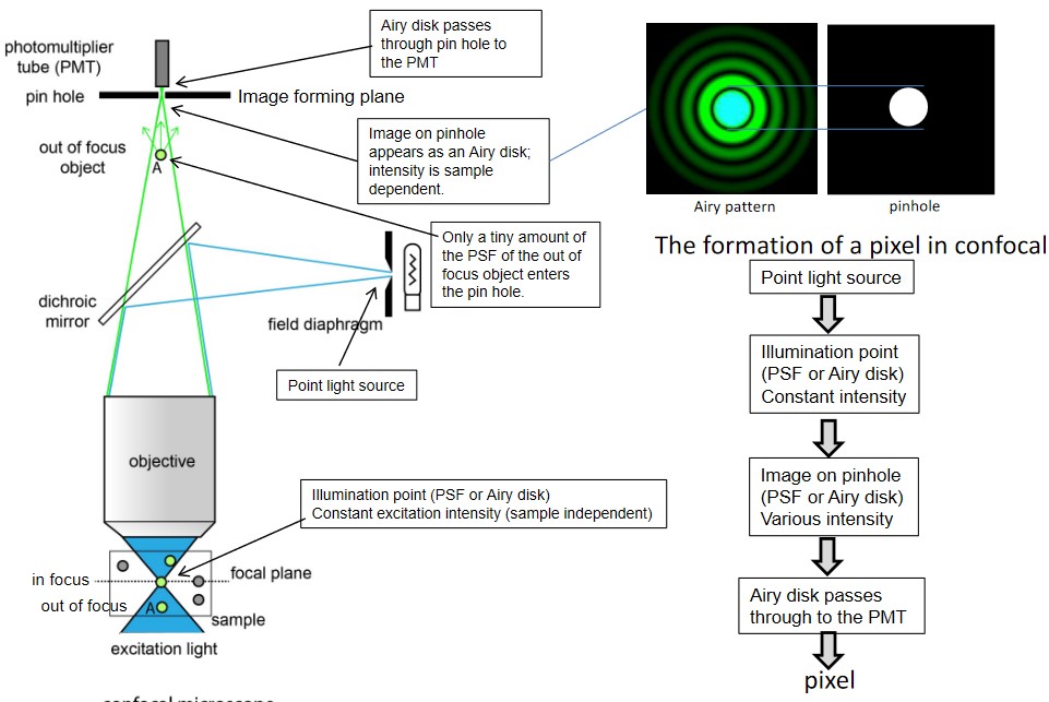
Figure 5.7: Confocal Microscopes Reducing Out-of-Focus Blurs
A point light source generates an illumination point that has constant intensity. This image on the pinhole then has various intensities - this image also passes through to the PMT. The pinhole is key as it removes out-of-focus light (there won’t be much energy lost as a result of this).
5.2.1 Scanning Mechanisms of Confocal Microscopes
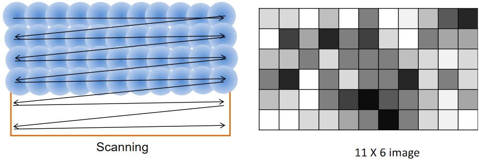
Figure 5.8: Image Scanning in Confocal Microscopy
Confocal microscopes use point-by-point scanning; each illumination point is the image of a point light source (i.e., a PSF or an airy disk).
The scanned point is then shown as a pixel in the final image.
5.2.2 Features of the Confocal Microscope
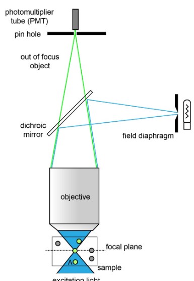
Figure 5.9: A Sample Confocal Microscope
A confocal microscope uses epi-fluorescence illumination and a laser as its excitation light source.
The pinhole at the top of the microscope removes out-of-focus blurs; a larger pinhole collects more light, but might suffer in confocality: the ability to remove out-of-focus blurs.
The photomultiplier tube (i.e., PT tube) doesn’t have spatial resolutions; confocal microscopes also use an xy-scanning system.
5.2.2.1 PT Tube
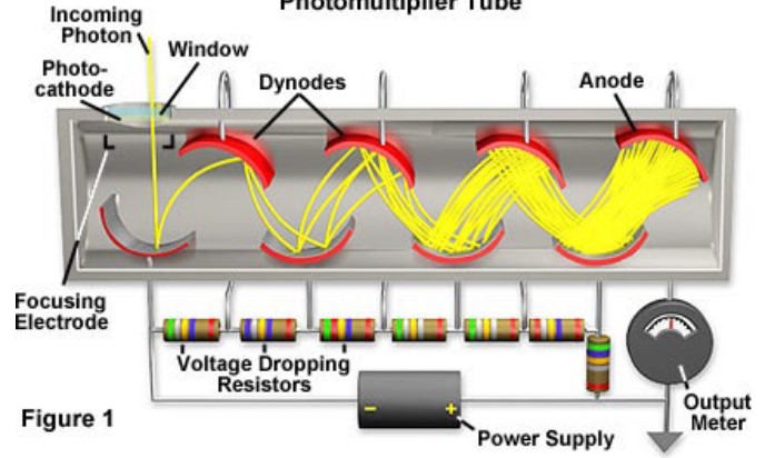
Figure 5.10: Schematic of a PT Tube
A photomultipler tube works as such:
- Photons strike the photocathode (which is made from photosensitive material), releasing photoelectrons in the process (this is called the photoelectric effect).
- The photoelectrons in 1. accelerated by the electric field imposed by dynodes.
- When electrons strike the dynode, more electrons are released.
- Steps 2. and 3. are repeated at various dynodes.
- Amplified electrons are collected by the anode.
5.2.3 Image Acquisition
The pixel dwelling time is measured in microseconds; the longer the dwelling time, the less the confocality of the microscope. It also uses laser power and PMT.
Image acquisition also needs a computer to process the pixels that are formed by aggregating pixels via software.
The black level is a parameter that can be used to truncate an image.