6.1 Approaches for Super-Resolution Microscopy
BS2010 covers two main approaches:
Engineered PSF
In this case, the PSF of the microscope is made smaller as is the case with Stimulated Emission Depletion (i.e., STED) microscopy.
Single Molecule Localization
The PSF remains the same, but the localization precision is increased by Gaussian fitting or adopting the center of mass as is the case with PALM and STORM.
6.1.1 Concepts Behind Single Molecule Localization Super-Resolution Microscopy
6.1.1.1 Gaussian Fitting
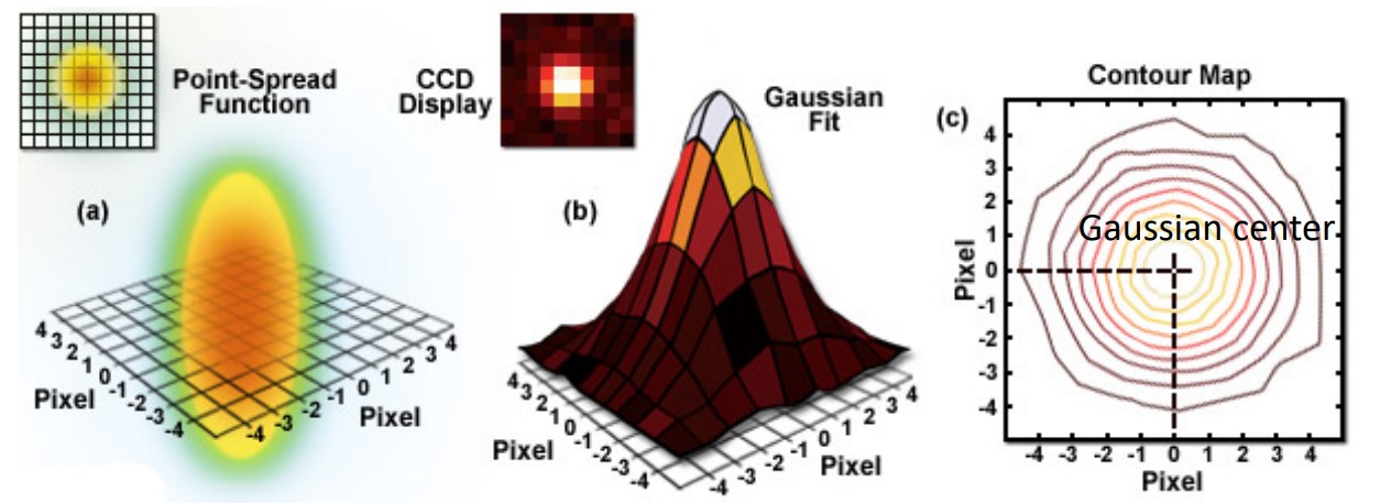
Figure 6.1: Gaussian Fitting
The image of a single molecule is Point Spread Function (i.e., PSF) in 3D coordinates or an Airy disk in 2D coordinates.
This airy disk can be fitted using a 2D Gaussian function. The Gaussian center is the position of the molecule at a nanometer resolution.
6.1.1.2 Center of Mass and Precision
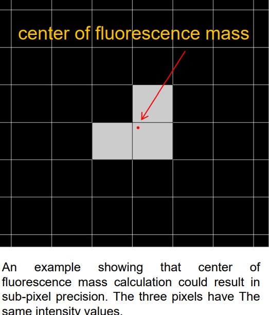
Figure 6.2: Center of Mass in Fluorescence Mass Calculation
The center of mass is an imaginary point where all the mass in an object is concentrated. If the PSF is a physical object whose mass is equivalent to its fluorescence intensity, then its center of mass is the location of the molecule.
The coordinates of the center of mass \(x_c\) are:
\[\begin{align} x_c = \frac{\sum_{i = 1}^nxi \times li}{\sum_{i = 1}^nli} & y_c = \frac{\sum_{i = 1}^nyi \times li}{\sum_{i = 1}^nli} \end{align}\]
Both the Gaussian center and center of mass give similar localization results with sub-pixel precision.
For single-molecule localization microscopy, its precision is represented by \(\sigma\) (i.e., standard deviation):
\[\begin{equation} \sigma = \frac{1}{\sqrt{n}} \end{equation}\]
Where \(n\) is the number of photons. As \(n \rightarrow \infty\), \(\sigma \rightarrow 0\)
6.1.2 Inner Workings of PALM and STORM
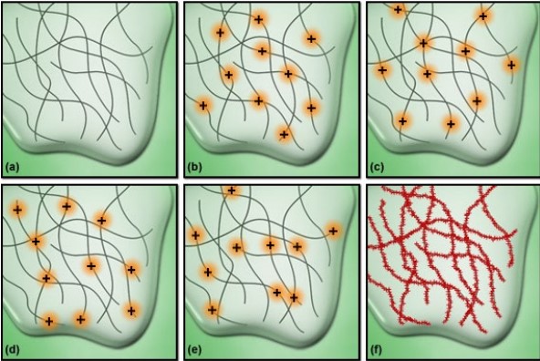
Figure 6.3: Super-Resolution Microscopy
PALM is short for photoactivated localization microscopy while STORM is short for stochastic optical reconstruction microscopy.
In both types of microscopy,photoactivatable (i.e., PA) fluorescent proteins and dyes are used; their properties can be activated by an activation light in the following steps:
- All fluorophores are initially dark and not activated.
- A random amount of fluorophores are turned on via an activation beam.
- Each molecule will be localized at tens of nanometer accuracy via Gaussian fitting.
- Each activated fluorophore will be turned off via photobleaching.
- Steps 2 to 4 are repeated numerous times.