Chapter 13 Inter-Organ Communications
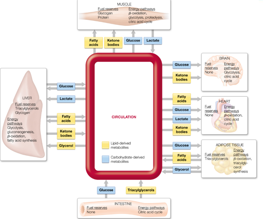
Figure 13.1: Metabolic Interactions in Major Organs
The major fuel metabolites imported and exported by each organ (and the major energy pathways that occur in each organ) is shown in the above figure.
All lipid-derived metabolites are highlighted in yellow; carbohydrate-derived metabolites are highlighted in blue.
The nervous system and the endocrine system are responsible for communication throughout the body.
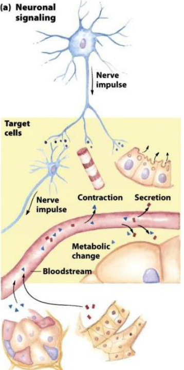
Figure 13.2: Neural Signalling
In neuronal signalling, electrical signals originate in the cell body (of a neuron) and travel a very long distance to the tip of the axon, where neurotransmitters (see below) are then released into the synapse and diffuse into the target cell.
In the endocrine system, hormones are secreted into the bloodstream - the bloodstream then carries the hormones throughout the body to target tissues that may be a meter or more away from the secreting cell.
13.1 Neurotransmitters and Neurohormones
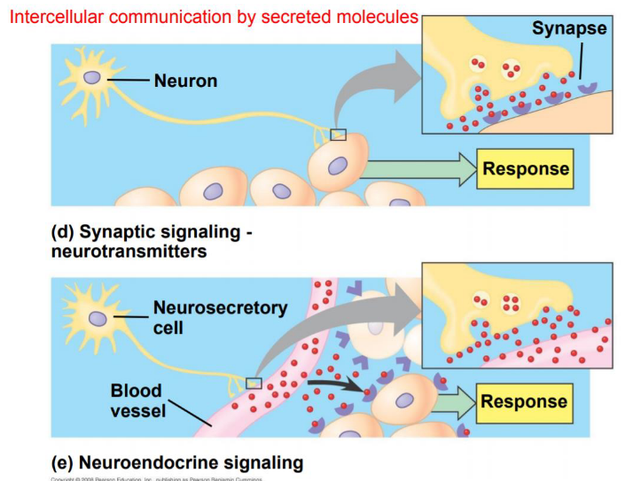
Figure 13.3: Neurohormones and Neurotransmitters
All neurons contact their target cells at synapses. There, chemicals signals called neurotransmitters diffuse a short distance to bind to the receptors on the target cell. All neurotransmitters also play a role in sensation, memory, cognition, and movement.
Neurohormones are a class of hormones that originate from the neurons in the brain and diffuse throughout the bloodstream.
13.2 Hormonal Regulation
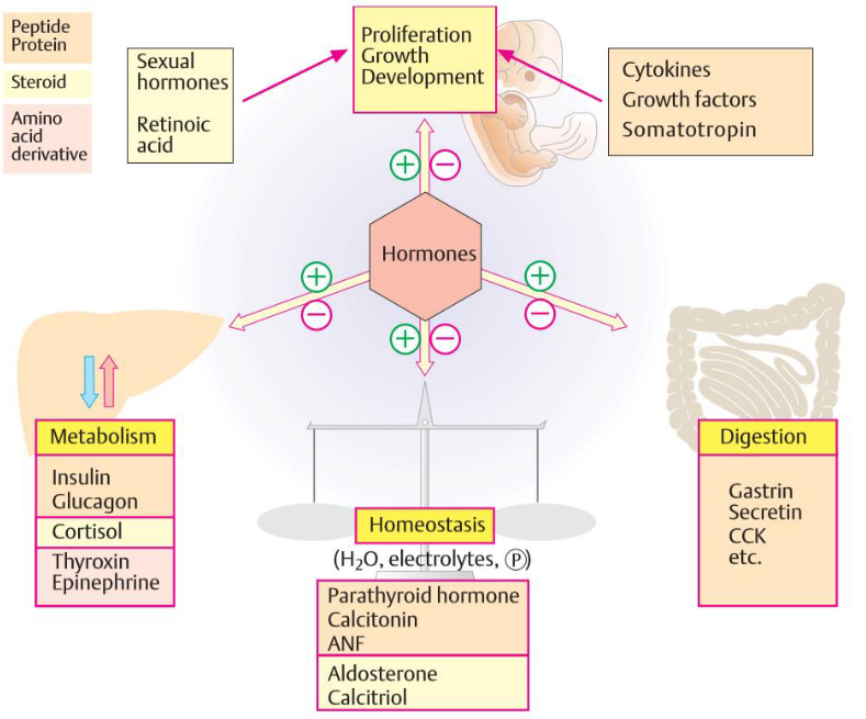
Figure 13.4: Hormonal Regulation
There are four components to the above graphic:
Growth and differentiation of cells, tissues, and organs
These include cell proliferation, sexual differentiation, and embryonic development. Steroid hormones are responsible for the latter via transcription regulation.
Metabolic pathways
These generally require rapidly-acting mechanisms; many hormones involved in metabolic pathways hence regulate the interconversion of enzymes.
Digestive processes
These are usually regulated by locally-acting peptides, but mediators, biogenic amines, and neuropeptides are also involved.
Maintenance of ion concentrations
In other words, homeostasis.
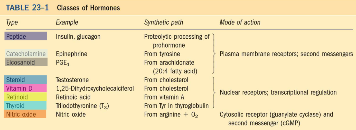
Figure 13.5: Classes of Hormones and their Modes of Action
Mammals also have several classes of hormones that differ by their chemical structures and their modes of action.
13.3 Cytokine Regulation
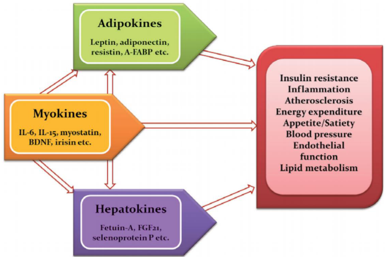
Figure 13.6: Some Cytokines
Cytokines are a large group of proteins that are secreted by specific cells of the immune system. Many signalling molecules produced by metabolic organs have been identified and named as:
- Hepatokines (liver)
- Myokines (muscle)
- Adipokines (white adipose tissue)
- Batokines (brown adipose tissue)
These cytokines can respond to variety of metabolic stimulants, including but not limited to fasting and feeding cycles, the circadian rhythm, exercise, and cold exposure.
13.3.1 Cytokine regulation of fuel metabolism
Cytokines can regulate cellular activity in a corrdinated, interactive way due to the following attributes:
- Pleiotropy - one cytokine has many different functions.
- Redundancy - several different cytokines mediate the same or similar functions.
- Synergism - the combined effect of two or more cytokines are greater than that of one.
- Antagonism - the effects of one cytokine offsets the effects of another cytokine.
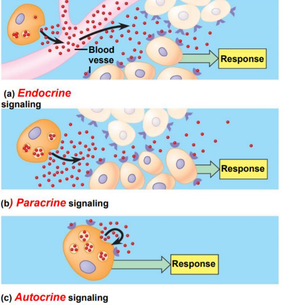
Figure 13.7: Types of Signals
Hormones and cytokines can be classified by the way they get from the point of their release to their target tissues:
- Endocrine hormones are released into the blood and carried to target cells throughout the body.
- Paracrine hormones are released into the extracellular space and diffuse into neighboring cells.
- Autocrine hormones are released by and affect the same cells - they also bind to receptors on the cell surface.
13.3.1.1 NB:
Hormones are a class of proteins that act as signalling molecules - these are secreted by a given tissue and are used to communicate between organs to trigger specific stimuli
Cytokines are a class of small proteins that can be produced by a broad variety of cells and act as signalling molecules. Cytokines are usually found in smaller concentrations than hormones.
13.4 Signal Transduction Pathways
Receptor activation happens when the messenger interacts with the receptor such that it induces a change in the conformation of the receptor - this is also the first step leading to the cell’s ultimate response.
Hence, the sequence of events between receptor activation and the responses that follow thereafter are called signal transduction pathways.
These response can also take the form of changes in:
- Metabolism
- Secretory activity
- Proliferation rate and differentiation
- Contractile activity
- Permeability, transport, and the electrical state of the cell’s plasma membrane
13.4.1 Lipophilic hormones
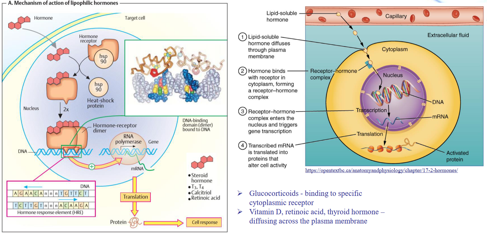
Figure 13.8: Mechanism of Lipophilic Hormones
Observe the above graphic for lipophilic hormones.
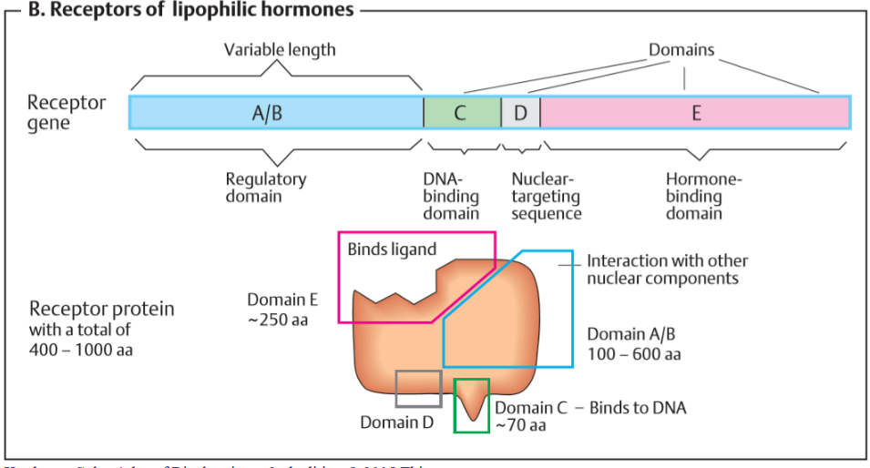
Figure 13.9: Receptors of Lipophilic Hormones
Also observe the following graphic.
13.4.2 Hydrophilic hormones
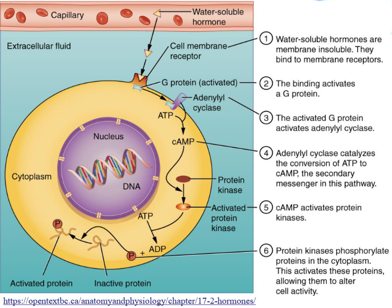
Figure 13.10: Mechanism of Hydrophilic Hormones
Observe the following graphic for the mechanism of a hydrophilic hormone.
13.4.2.1 1-helix receptors
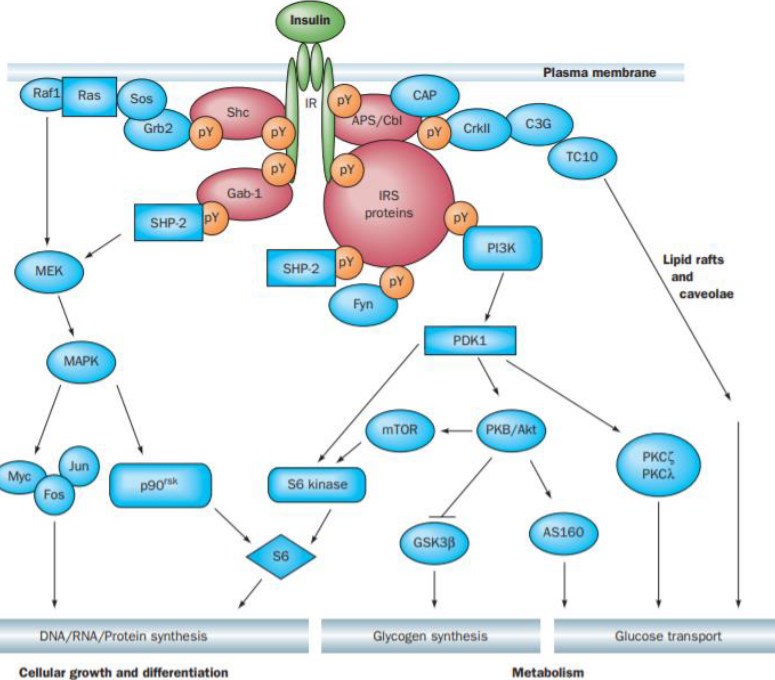
Figure 13.11: Insulin Signalling System
While the above signalling system isn’t fully understood, it is obviously very complex.
Upon binding to insulin, the insulin receptor autophosphorylates itself at several tyrosine residues and then phosphorylates its own proteins, hence activating several signalling pathways that control a diverse array of effects.
Hence, a hormone like insulin can trigger many different effects that wouldn’t be possible in one hormone.
13.4.2.2 7-helix receptors
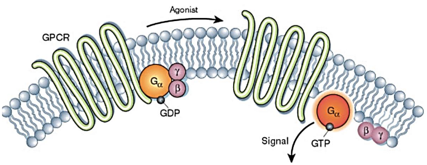
Figure 13.12: 7-Helix Receptors
GCPR proteins can be grouped into six classes (A to F) based on sequence homology and functional similarity.
Glucagone are class B hormones; epinephrine acts on \(\beta\)-adrenergic receptors and \(\alpha2\)-adrenergic receptors.
13.4.2.3 G-proteins signal transduction
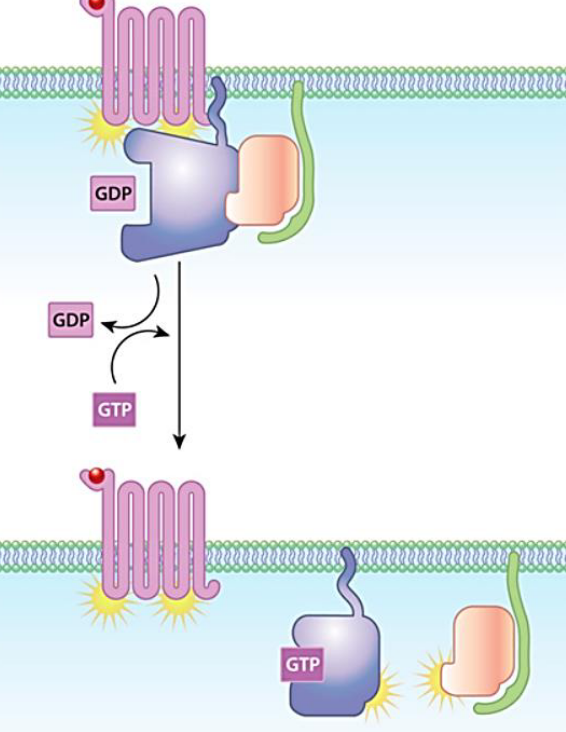
Figure 13.13: G Protein Signal Transduction
Observe the following graphic above.
13.5 Different Types of Second Messengers
The concentration of these intracellular messengers are regulated by hormones, neurotransmitters, and other extracellular signals. They are also made from easily available substrates and have a short half-life:
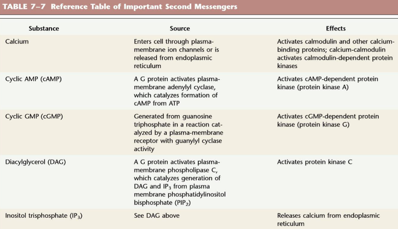
Figure 13.14: Sources and Effects of Second Messengers
- cAMP
- cGMP
- Ca2+
- Inositol triphosphate (InsP3)
- Diacylglycerol (DAG)
- Nitrogen monoxide (NO)
13.5.1 Mechanism of cyclic AMP
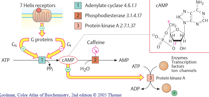
Figure 13.15: cAMP Action
cAMP is made from membrane-bound adenylate cyclases, the degradation of which to AMP is catalyzed by phosphodiesterases (inhibited by caffeine).
Adenylate cyclase activity is regulated by G proteins (Gs and Gi).
13.5.2 Mechanism of Ca2+ ions
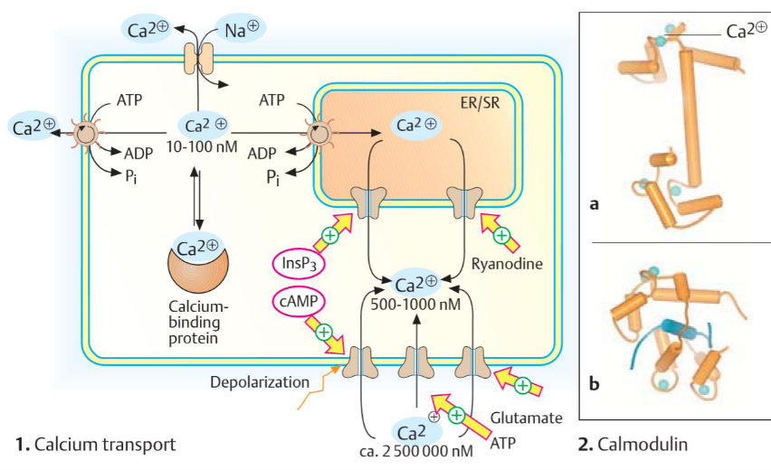
Figure 13.16: Ca2+ Ion Action
Ca2+ is a signalling ion - its concentration in the cytoplasm is very low (10 nM - 100 nM).
Specific signals can trigger a sudden increase in Ca2+ levels to 500 nM to 1000 nM by opening Ca2+ channels in the plasma membrane or the membranes of the endoplasmic reticulum or the sarcoplasmic reticulum.
13.5.3 Mechanism of InsP3 and DAG
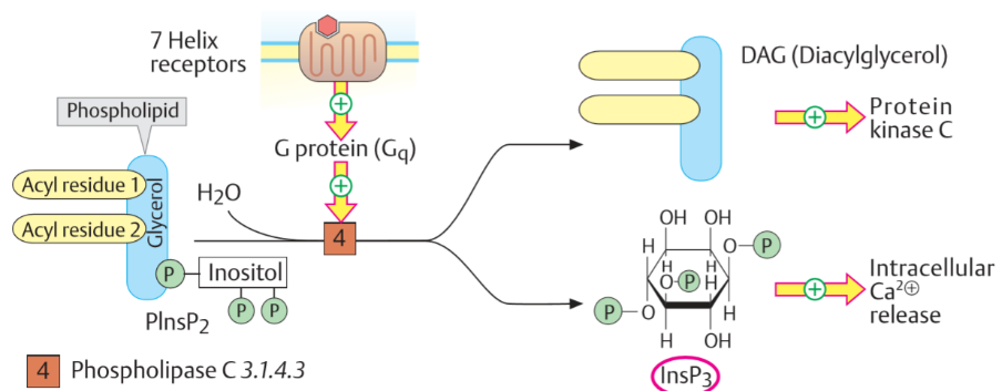
Figure 13.17: InsP3 and DAG Action
Gq proteins activate phospholipase C (i.e., PLC). PLC then creates two messengers: inositol 1,4,5-triphosphate phosphate and diacylglycerol.
InsP3 migrates to the endoplasmic reticulum and opens Ca2+ ions that allow Ca2+ ions to flow into the cytoplasm.
13.6 Regulation of Hormonal Release
The central nervous system receives inputs from many internal and external signals and produces the appropriate hormone signals via the endocrine system.
The hypothalamus is the coordination center of the endocrine system.
The pituitary gland has two functional units:
- The posterior pituitary has the axonal endings of many neurons that originate in the hypothalamus.
- The anterior pituitary responds to hypothalamic hormones in the blood; tropins and tropic hormones are produced.
13.6.1 Hormone cascades
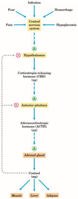
Figure 13.18: Cascade of Hormones
At each step of the cascade, feedback inhibition of earlier steps in the cascade is possible. An unnecessarily high level of the ultimate hormone or of one of the intermediates inhibit the release of the earlier hormones in the cascade.
These feedback mechanisms accomplish the same end as those that limit the output of the biosynthetic pathway: a product is made (or released) until the necessary concentration is reached.