Chapter 5 Evolution and Taxonomy
Microorganisms (and organisms in general) can evolve via a multitude of ways, including but not limited to:
- Drift (i.e., diversification)
- Selection (fixation and loss)
- Heritable changes in the organism’s genetic code
- This can include mutations or chromosomal rearrangments.
- Horizontal gene transfers may also occur. These include plasmids, transposons, DNA uptake and recombination, viruses, and bacteriophages.
5.1 What is a Species?
“The species… is the basic unit of ecology… no ecosystem can be fully understood until it has been dissected into its component species and until the mutual interactions of these species are understood..”
— Ernst Mayr (Zoologist)
However, as Mayr himself put it, a species is:
“[a] [group] of interbreeding or potentially interbreeding natural populations that are reproductively isolated from other such groups”
— Ernst Mayr (Zoologist)
Yet, it is important to realize that prokaryotic species are very similar to one another. Hence, prokaryotic strains are to be allocated as “species” when:
“…they are at least approximately 70% identical base pair sequence (DNA similarity) and a difference in the melting point [Tm_e] of DNA/DNA homoduplexes and heteroduplexes of less than 5°C…”
— Bergey’s Manual
NB: Bergey’s manual is more taxonomic than it is phylogenetic!
Note that genomospecies are not to be named unless they can be differentiated from other species on a known basis of some phenotypic feature and form a monophyletic group.
5.1.1 Why might one choose to study evolutionary data?
For one, doing so may enable us to understand how and when bacteria acquired antibiotic resistance.
Furthermore, studying evolutionary data could let us better other organisms’ relatedness to each other (including humans).
Evolution, as it relates to organisms, has more to do with its members’ growth rate and yield. Nutrient capture processes are needed acquire energy for reproduction and cellular processes.
5.1.2 Levels of taxonomic classification
Taxonomy is a branch of science that deals with the classification of living organisms.
The levels are (in order of greatest to lowest):
- Domain
- Kingdom
- Phylum
- Class
- Order
- Family
- Genus
- Species
5.1.3 Morphospecies
A morphospecies is a species based on numerical taxonomy: a battery of biochemical tests that are each assigned a positive or a negative value.
A battery of 300 tests can reveal as litle as 5 - 20% of the genetic potential of a strain of bacteria!
Lateral gene transfer between bacteria also confuse results!
5.1.3.1 DNA-DNA hybridization (DDH)
This method is considered the “gold” standard for species identification.
This method relies on DNA (obviously), provides genomic information about a certain kind of bacteria, and are more discriminatory than phenotypic tests.
However, such a method is often tedious and difficult to replicate.
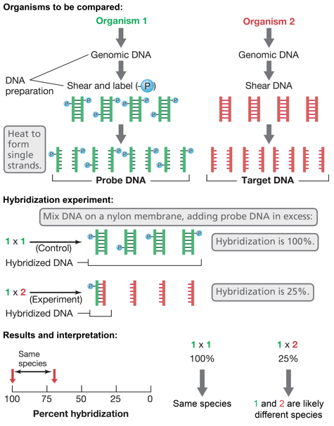
Figure 5.1: DNA-DNA Hybridization Process
5.1.3.2 16s rRNA: the “gold standard” phylogenetic marker?
One could identify a species of bacteria by sequencing its 16s rRNA.
Indeed, such a method could be ubiquitous in prokaryotes, not to mention the availability of universal primers and the fact that variable regions can be used to compare closely related species.
However, such a comparison does not represent the bacteria’s entire genome and may result in a poor resolution in some species and genera. There is also evidence of horizontal gene transfer between microbes, and species can exist as multiple copies.
A 16s sequence identity of less than 97% between strains indicate that both bacterial strains are different species. At anything greater than or equal to 97%, DNA-DNA hybridization must be used.
5.1.4 NB…
There is no official species concept for prokaryotes, and there is no official classification of bacteria and archaea.
Instead, a polyphasic taxonomy is used:
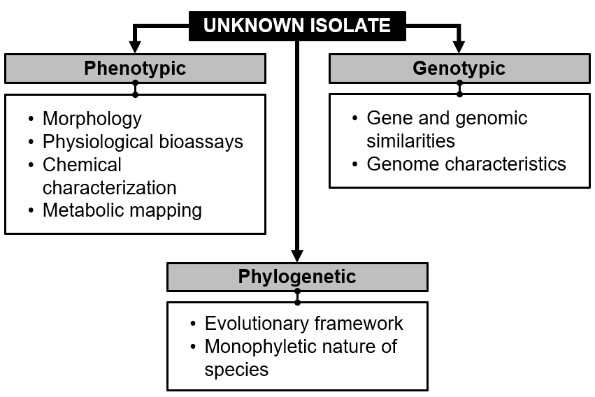
Figure 5.2: A Polyphasic Approach
5.2 Uncultured Diveristy
5.2.1 The great plate count anomaly
Direct counts of bacteria in natural samples are typically much higher (~1000 times more) than those from viable plate counts.
Syntrophy is a phenomenon where bacteria depend on other bacteria to survive.
However, bacteria can be in different stages of life in plate counts. They could be dead, virally infected and unable to form colonies, viable, but nonculturable, or are culturable, but the right food isn’t given.
The major implication? We don’t know much about more than 99% of all prokaryotes!
5.2.2 Culture and uncultured diversity
There are 92 bacterial phyla, 26 archaeal phyla, and 5 eukaryotic supergroups:
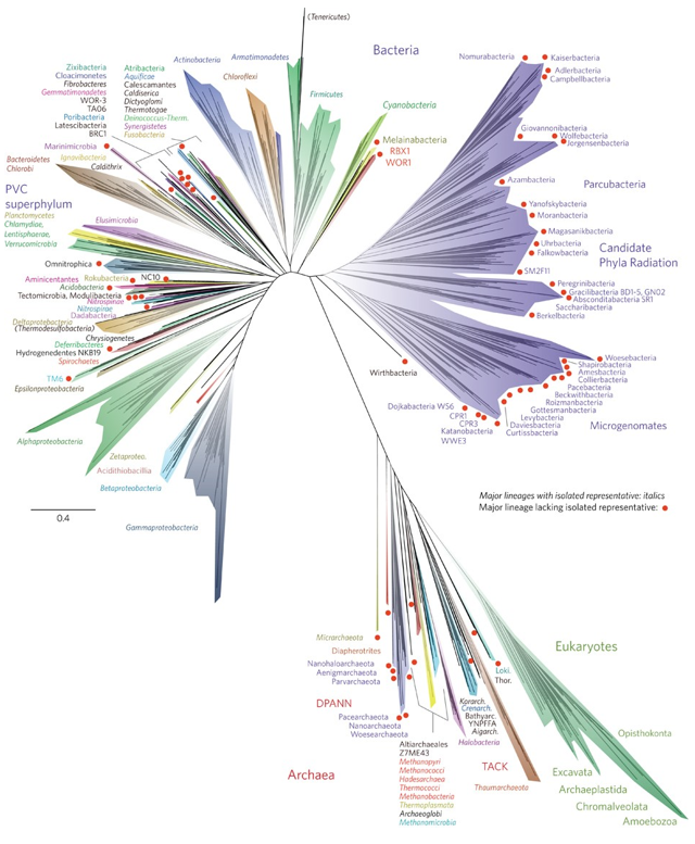
Figure 5.3: Phyla of Three Domains of Life
The red dots above represent lineages that lack an isolated representative.
5.3 Firmicutes (i.e., gram positive bacteria)
5.3.1 Bacillus
Bacillus shapes occur when bacteria age due to nutrient deprivation. This is not triggered by environmental stress and is extremely resistant to environmental stresses.
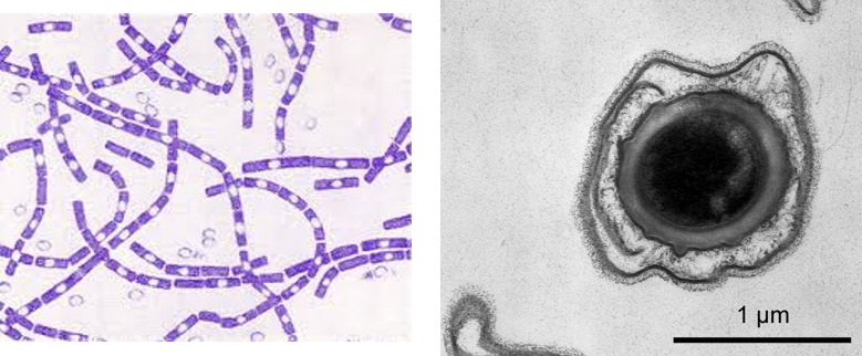
Figure 5.4: Bacillus Bacteria
5.3.2 Streptomyces
These are filamentous actinobacteria that have linear chromosomes.
They are also the source of antibiotics used today:
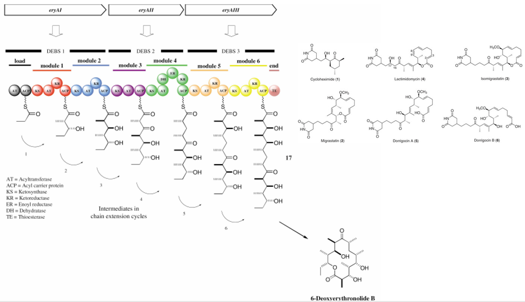
Figure 5.5: Antibiotic Production in Streptomyces Bacterium
Interestingly enough, Streptomyces bacteria also grow “roots” into the nutrient agar (hence, when cultivated, one must dig them out) and form spores in these roots.
5.3.3 Paenibacillus
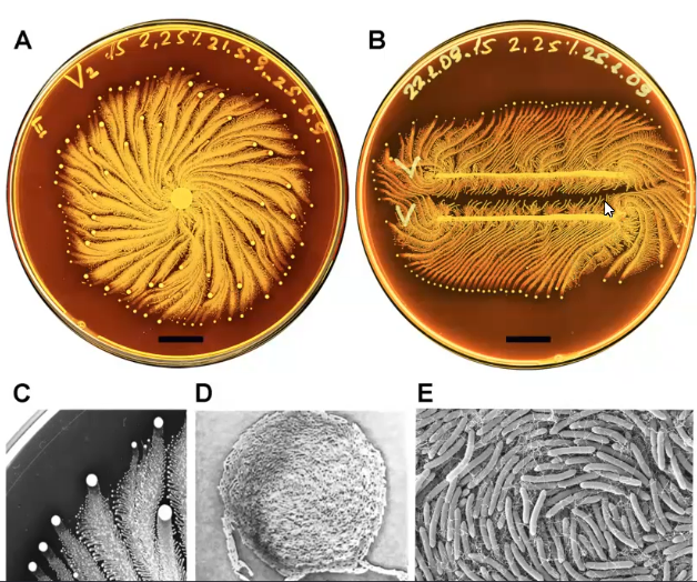
Figure 5.6: Paenibacillus Pictures
These are bacteria whose motilities aren’t quite understood. These bacteria form large spiral motions and are somehow able to “sense” each other.
5.4 Free-Living Microbes
5.4.1 Pelagibacter ubique
These are bacteria that were discovered in the Sargasso sea. They make up 50% of cells in temperate waters and are photosynthetic.
These bacteria also have the smallest genomes among free-living organisms: 1.3 Mb in total!
5.4.2 Myxobacteria
These bacteria have some of the largest genomes (e.g., 13 Mb) and displays cellular differentiation (including but not limited to fruiting bodies):
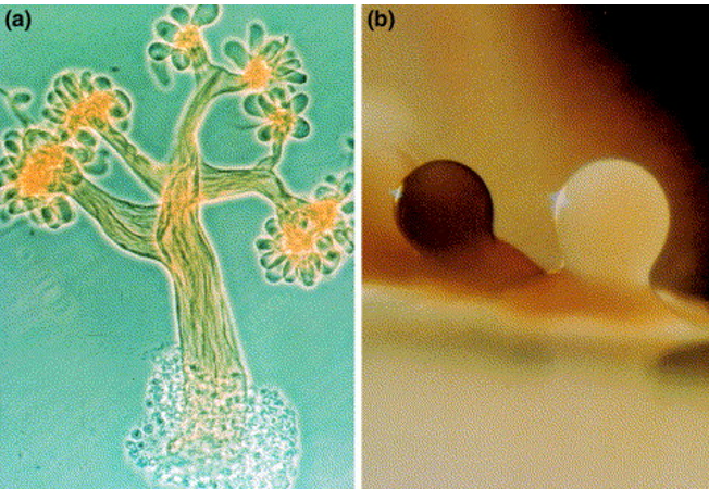
Figure 5.7: Fruiting Bodies in Myxobacteria
Furthermore, Myxobacteria are able to remotely sense objects and hunt down prey in a coordinated attack.
5.4.3 Caulobacter crescentus
These bacteria divide asymmetrically:
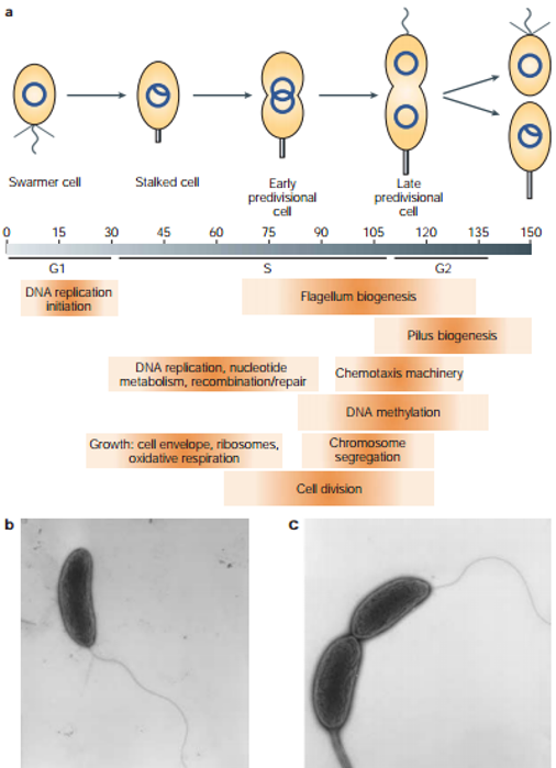
Figure 5.8: Asymetrical Division of Caulobacter crescentus
As mentioned in chapter 3 of this website, caulobacter is also used as a model organism for studying complex prokaryotic cell processes (especially cellular division)!
5.4.4 Denicoccus reidurans
This is a bacterium that was isolated from a nuclear reactor. It is able to withstand up to 10000 Grays (5 Grays is enough to kill a human in two weeks), is highly pigmented (with carotenoid pigments that protect against UV irradiation), has more than one chromosomes, has a highly active DNA repair system, and has mechanisms to protect DNA-repairing proteins.
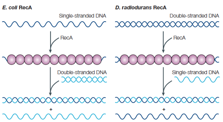
Figure 5.9: DNA Exchange in E. coli and D. reidurans
5.4.5 Helicobacter pylori
This bacterium was discovered in 1982 by Dr. Barry Marshall and Dr. Robin Warren of Perth, Western Australia in the stomachs of patients with gastric ulcers.
H. pylori performs chemotaxis through mucus towards the epithelial cells to avoid the high acidic environment in the stomach.
The bacterium’s pathogenicity is due to the Cag pathogenicity island: an island that encodes for several virulence determinants, including the type IV secretion system and the cagA gene (which encodes for a protein that disrupts the host cell’s activity).
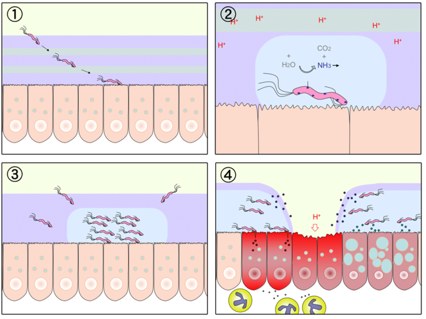
Figure 5.10: H. pylori Causing Ulcers
5.4.6 Bdellivibrio
This is a bacterium that attacks other gram negative bacterium by colliding with them at speeds up to 160 \(\mu\)m per second - 100 times their length per second using a single sheathed polar flagellum for movement with a dampened filament waveform for movement.
The bacterium then attaches itself to the host cell’s outer membrane and peptidoglycan layer, after which it creates a small hole in the outer membrane.
The bacterium then enters the host cell via the host’s periplasmic space, after which it remains attached to it for a brief “recognition” period. The bacterium then becomes irreversibly attached via the pole opposite the flagellum before sealing the aforementioned hole and converting the host cell into a spheroblast.
A mixture of hydrolytic enzymes is applied in a manner that minimizes damage to the host cell - the two cell complex is called a bdelloplast.
Nevertheless, the Bdellovibrio bacterium then uses hydrolytic enzymes to degrade the host cell’s molecules to elongate and form a filament. The filament then seprates to form Bdellivibrio offspring.
The offspring then cause the host cell to lyse and the offspring to be released into the environment. The entire life cycle takes about three hours and produces an average of 3 - 6 offspring per E. coli bacterium.
5.4.7 Heterocysts (of cyanobacteria)
Heterocysts have several features in place for nitrogen fixation:
- They have thickened cell walls to reduce cell permeability
- They lack chlorophyll (and hence are unable to undergo photosynthesis)
- Reduced nitrogen is transported from the heterocyst to vegetative cells.
- Conversely, sugar is transported from vegetative cells to heterocysts.
- Sugars are supplied in excess in the event that they leak out of the cell, heterotrophic bacteria are attracted and consume both oxygen and sugars reducing oxygen.
- The diffusion of these compounds determines heterocyst spacing.
- Sugars are supplied in excess in the event that they leak out of the cell, heterotrophic bacteria are attracted and consume both oxygen and sugars reducing oxygen.
- Conversely, sugar is transported from vegetative cells to heterocysts.
5.5 Obligate Symbionts
5.5.1 Buchnera aphidicola
These bacteria belong to the gammaproteobacteria.
These bacteria also live in specialized aphid cells, are unable to repair DNA, has no lipopolysaccharides, and overproduces amino acids.
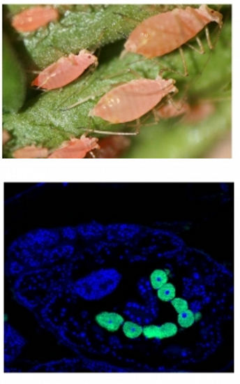
Figure 5.11: Aphids and Buchnera aphidicola
5.5.2 Mycoplasma and Phytoplasma
Mycoplasma and Phytoplasma are microbes that infect animals and plants respectively. Mycoplasma causes tuberculosis in humans!
In 1962, R.G.E. Murray proposed to divide the bacterial kingdom into three divisions based on the basis of cell wall types:
- Gram negative “Gracilicutes”: a thin cell wall and little peptidoglycan
- Gram positive “Firmacutes”: a thicker cell wall and more peptidoglycan.
- “Mollicutes”: those without a cell wall.
5.6 Origin of Bacterial Organelles
Mitochondria arose as a result of a eukaryotic ancestor engulfing a gamma proteobacterium (i.e., endosymbiosis).
Chloroplasts result from a single event: an Archaean engulfing a cyanobacterium - the engulfed cyanobacterium then became a plastid.
Plastids are organellles that are responsible for processes like photosynthesis, pigment synthesis, and storage of products like starch.
Both plastids and mitochondria divide by binary fission (like bacteria).