Chapter 9 Lesion-symptom mapping
Written by Vitória Piai
Reviewed by Brielle Stark
As we saw in Section 6, neurological conditions can lead to language production deficits or aphasia. To identify the most critical brain areas associated with language production, lesion-symptom mapping (LSM) techniques can be employed (Haan and Karnath 2018). Note that these techniques can be applied to virtually any type of behavioural variable.
One commonly used technique is so-called voxel-based lesion-symptom mapping (VLSM) (Bates et al. 2003). Using individuals’ behavioural scores (e.g., naming accuracy) and a delineation of their affected brain area (e.g., Figure 9.1), a statistical test is run at the group level at every voxel, comparing scores between individuals with and without damage in that voxel. The resulting map depicts the voxels that are critical for the measured behavioural performance, allowing one to infer a casual relationship between a certain area and a behavioural deficit. Although other types of LSM techniques may work somewhat differently under the hood (e.g., based on volume), they all allow similar inferences, as you will see below.
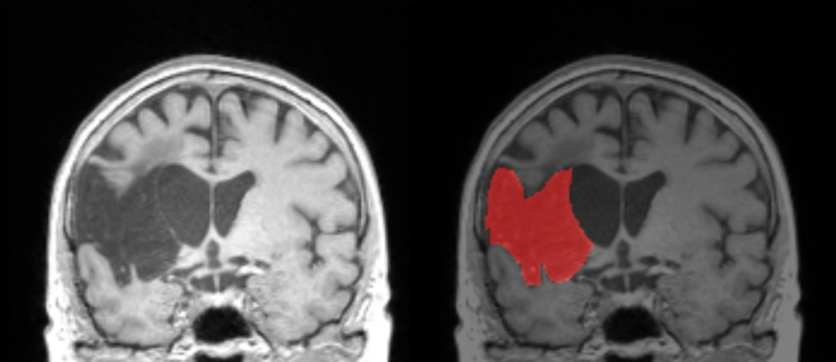
Figure 9.1: Illustration of a structural image with a stroke lesion (left) and the corresponding lesion mask in red (right). The lesion mask is expressed as a 3D volume of values. In this example, the voxels in red have the value of 1, that is, lesioned, whereas the non-coloured voxels have the value of 0, that is, not lesioned.
9.1 Evidence from stroke
A study examining picture naming in people with stroke in the chronic phase found that the mid and posterior portions of the left middle temporal gyrus (MTG) were critical for naming performance (Baldo et al. 2013). Figure 9.2 depicts these results, with the colours indicating the strength of this association.
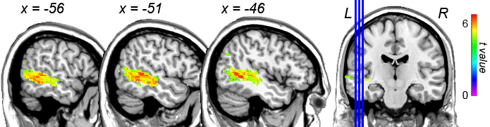
Figure 9.2: VLSM results for picture naming accuracy after controlling for visual and motor speech impairments. The colours indicate the strength of the associations. Only the left hemisphere is shown. L = Left; R = Right. VLSM results for making this figure are a courtesy of Juliana Baldo.
Using a measure of word error (i.e., paraphasias) derived from connected speech (i.e., whole sentences) in a picture description task, semantic paraphasias (real words that are semantically related to their intended targets, see Section 6) were related predominantly to the mid portion of the left superior (STG) and MTG. By contrast, unrelated paraphasias (real words that were semantically unrelated to their intended targets) were associated predominantly with posterior left MTG and inferior temporal gyrus (Stark et al. 2019). These results are shown in Figure 9.3. Finally, neologistic errors (non-words with no phonological relationship to their target) were associated with pre- and postcentral gyri and supramarginal gyrus. Also using semi-spontaneous speech, impairments in articulation and prosody were found in relation to damage in a frontal region comprising the left insula, the cortical structure that covers it (i.e., “operculum”), and a deeper structure called putamen (Henseler, Regenbrecht, and Obrig 2014). The role of the insula in articulation has also been suggested by a study that used a repetition task of words requiring complex articulatory gestures (Baldo et al. 2011).
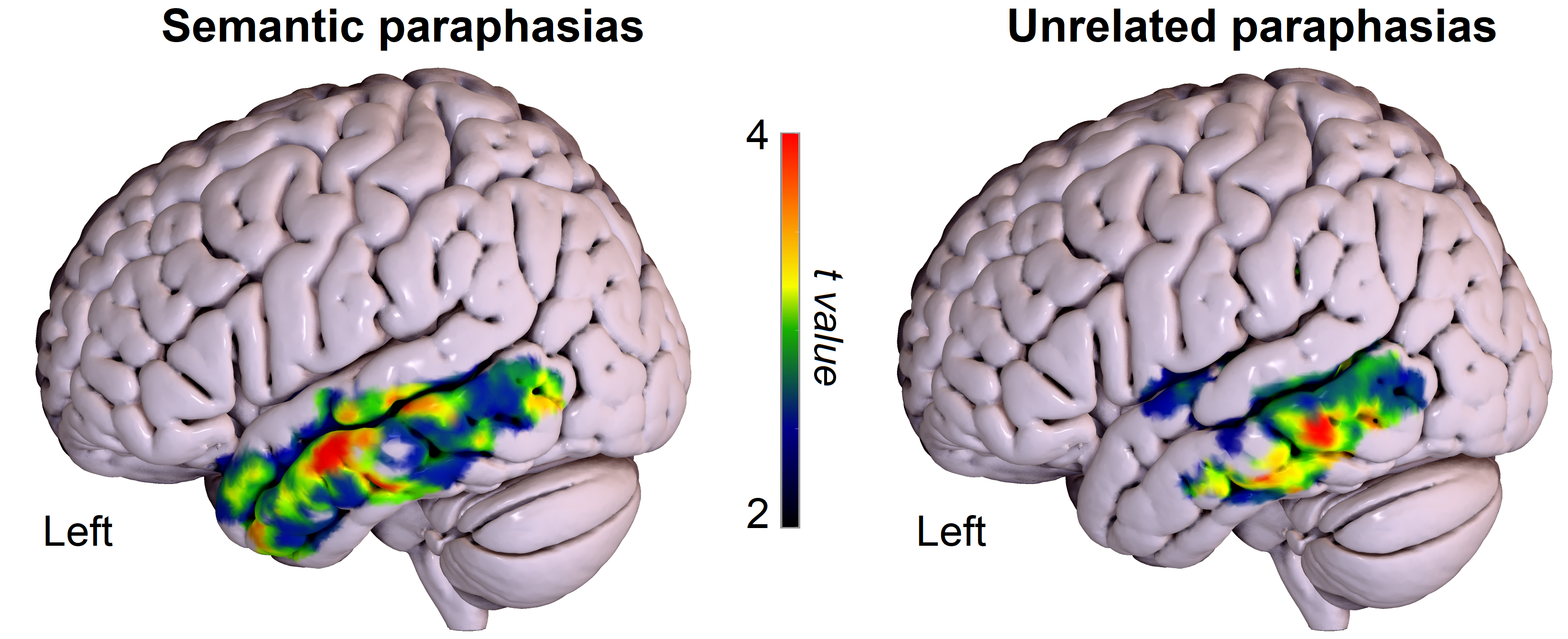
Figure 9.3: VLSM results for semantic (left) and unrelated (right) paraphasias. The colours indicate the strength of the associations. Only the left hemisphere is shown. This figure is a courtesy of Brielle Stark.
9.2 Evidence from brain tumour
In a sample of people with brain tumours prior to surgery, semantic paraphasias and omissions in picture naming were associated with tumours in the left posterior portion of the MTG (Faulkner and Wilshire 2020), while deficits in naming nonliving objects were particularly associated with tumours in the mid portion of the left MTG (Campanella et al. 2010; see also Fekonja et al. 2021; Pisoni et al. 2018). Impairment in phonological encoding were associated with tumours predominantly in the left posterior supramarginal gyrus and angular gyrus. Finally, articulatory-motor planning deficits were associated with the left frontal cortex, including the inferior frontal gyrus and the insula (Faulkner and Wilshire 2020).
Post-operatively, overall naming deficits two to three days after surgery were more likely when the mid portion of the left MTG was removed, and at one month post-surgery, an association still existed between left anterior MTG and naming deficits (Wilson et al. 2015).
9.3 Evidence from dementia
In primary progressive aphasia (which can occur as a result of several neurodegenerative disorders, including fronto-temporal dementia and Alzheimer’s disease), atrophy in the mid portion of the left temporal lobe was associated with the production of nouns of increasing lexical frequency (i.e., easier to produce) in connected speech, whereas phonological paraphasias were associated with atrophy in the mid portion of left STG. Finally, reduced speech rate was associated with atrophy of the left precentral gyrus and left inferior frontal gyrus (Wilson et al. 2010).
Take-home messages
- There is a large body of converging evidence across different neurological conditions for word production processes and certain brain areas
- Performance measures tapping into the conceptual and lexical stages tend to be associated with anterior-to-mid portions of the left temporal lobe, whereas phonological errors tend to be related to posterior temporal lobe, projecting dorsally to supramarginal gyrus and pre- and post-central gyri. There are however exceptions to this strict division
- Performance measures tapping into motor speech, such as articulation, are associated mostly with left frontal areas, both laterally and in structures lying underneath the lateral cortex and deeper
Suggestions for further reading
The interested reader is referred to additional literature on LSM, for example for measures derived from connected speech (Borovsky et al. 2007), for sentence production (Lukic 2021), for aphasia clinical measures (Fridriksson et al. 2018) and for a comparison between two LSM methods (Geva et al. 2012)